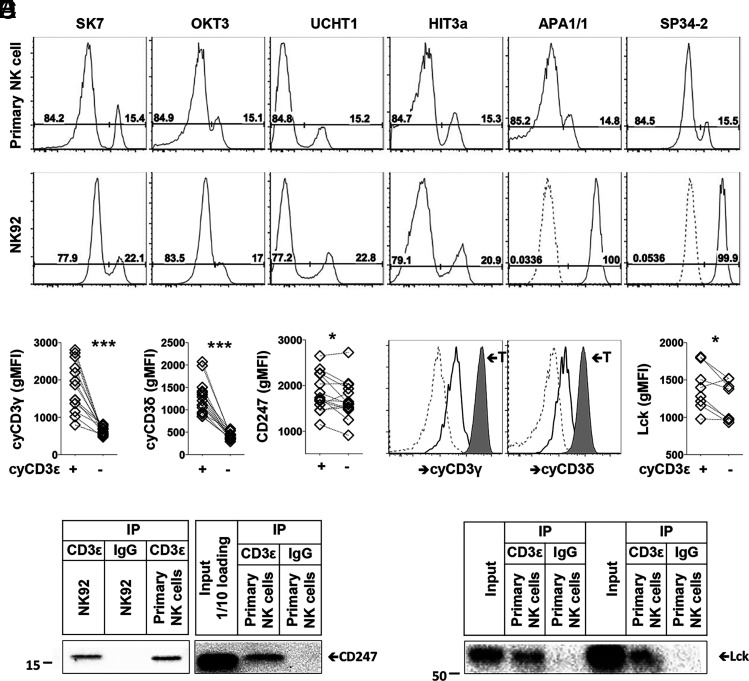
FIGURE 6.
CD3 subunits form complexes in HCMV-induced NK cells. (A) Detection of cyCD3ε+ NK cells using indicated CD3ε Ab clones among primary CD56dim NK cells and NK92 cells is shown. Isotype controls for APA1/1 and SP34-2 are indicated as dashed lines. (B) gMFIs of CD3γ, CD3δ, and CD247 on cyCD3ε+ and cyCD3ε– NK cells are shown. Populations from the same donors are paired (n = 14). (C) Expression of cyCD3γ and cyCD3δ is shown on cyCD3ε− NK cells (dashed), cyCD3ε+ NK cells (solid), and T cells (tinted) from the same donor. One representative donor out of 14 analyzed is shown. (D) NK92 cells and purified NK cells from two cyCD3ε+ NK cell donors were lysed, immunoprecipitated with control IgG or anti-CD3ε Ab and blotted with anti-CD247. (E) gMFIs of Lck on cyCD3ε+ and cyCD3ε– NK cells are shown. Populations from the same donors are paired (n = 8). (F) Purified NK cells from two cyCD3ε+ NK cell donors were lysed and immunoprecipitated with control IgG or anti-CD3ε Ab for Lck detection. *p < 0.05, ***p < 0.001.

