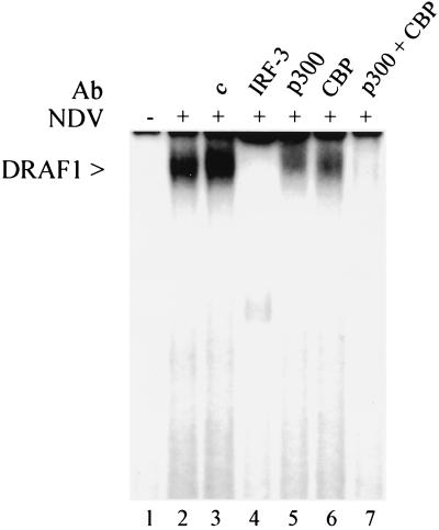
FIG. 1.
Appearance of DRAF1 in the nucleus following viral infection. Electrophoretic mobility shift assay was performed with nuclear extracts from HEC-1B cells either uninfected (lane 1) or infected with NDV for 6 h (lanes 2 to 7). The following specific antibodies were included in the DNA binding reactions: lane 3, 1 μg of control rabbit serum; lane 4, 1 μg of anti-IRF-3 antibody; lane 5, 1 μg of anti-p300 antibody; lane 6, 1 μg of anti-CBP antibody; and lane 7, 0.5 μg of anti-CBP antibody and 0.5 μg of anti-p300 antibody.

