Figure 2.
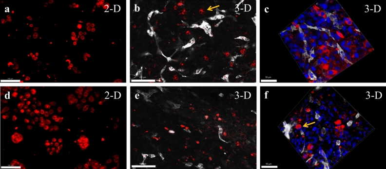
Formation of vascularized 3-D models with different breast cancer cell types. (a) Morphology of non-triple negative MCF-7 BCCs (red) in 2-D cell culture plate, Scale = 100 µm, (b) morphology of MCF-7 BCCs embedded in scaffold-free vascularized tissue, vessels were stained (grey) with PECAM1 (CD-31), arrow indicates grouping of MCF-7 cells that forms tumor clusters, Scale = 100 µm, (c) 3-D z-stacked image of MCF-7 within a vascularized tissue highlights its morphology, Scale = 50 µm, (d) morphology of triple-negative MDA-MB468 (red in 2-D culture, Scale = 100 µm, (e) MDA-MB468 display isolated and rounded morphology within scaffold-free vascularized models, (f) 3-D z-stacked image of MDA-MB468 and the vascularized 3-D tissue, arrow highlights the rounded morphology within the vascularized model, Scale = 50 µm.

