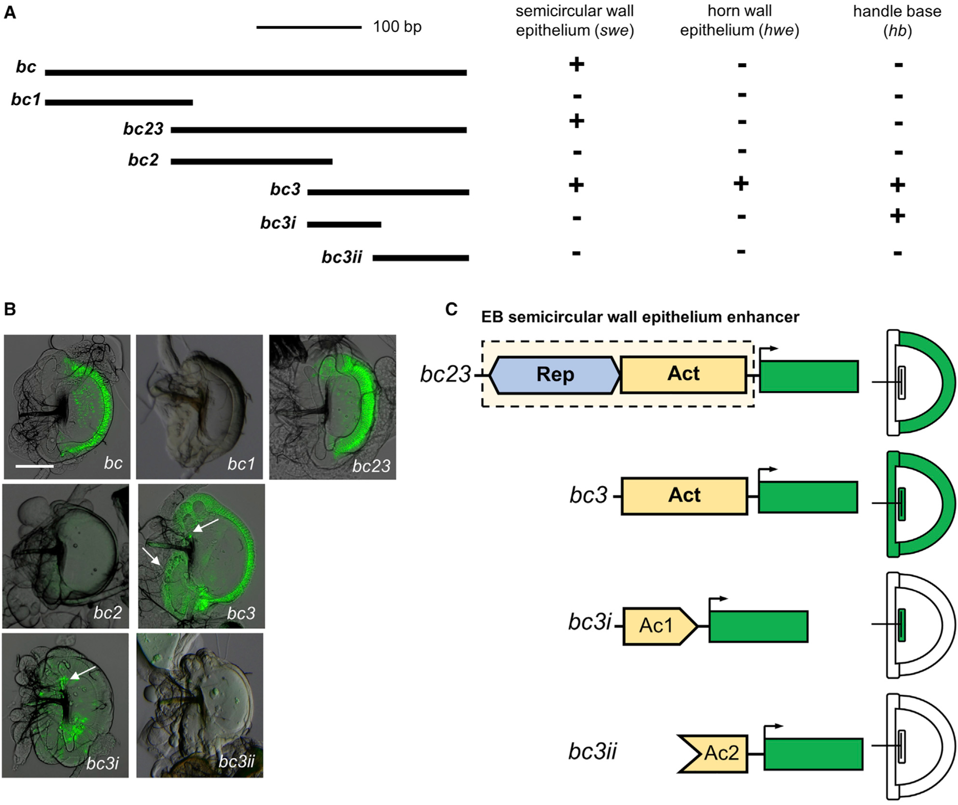
Figure 3. The combination of repressor and activator sequences in the D. melanogaster bond EB enhancer drives specific expression in the swe.

(A) Schematic of the overlapping GFP constructs of the EB enhancer. The bc23 fragment is the minimum region that can recapitulate the EB expression of bond in the swe. The bc3 fragment shows the ectopic GFP expression in horn wall epithelium (hwe) and handle base (hb). Plus sign (+) indicates the presence of expression; minus sign (−) indicates the absence of detectable expression.
(B) GFP reporter protein expression in the EB corresponding to the different overlapping constructs. Arrows indicate the expression in hwe and hb. Scale bar, 100 μm.
(C) The D. melanogaster bond EB swe enhancer contains two modular regions. The activator region (Act) contains activator sequences that drive expression in the whole EB (in hwe, hb, and swe). The repressor region (Rep) represses GFP activity in the hb and hwe of EB and restricts activity to the swe of the EB. The Act region can be divided into two different inputs, Ac1 and Ac2, both of which are needed to drive expression in the whole EB. Ac1 on its own can drive GFP expression in the hb, but Ac2 alone does not drive any expression.
