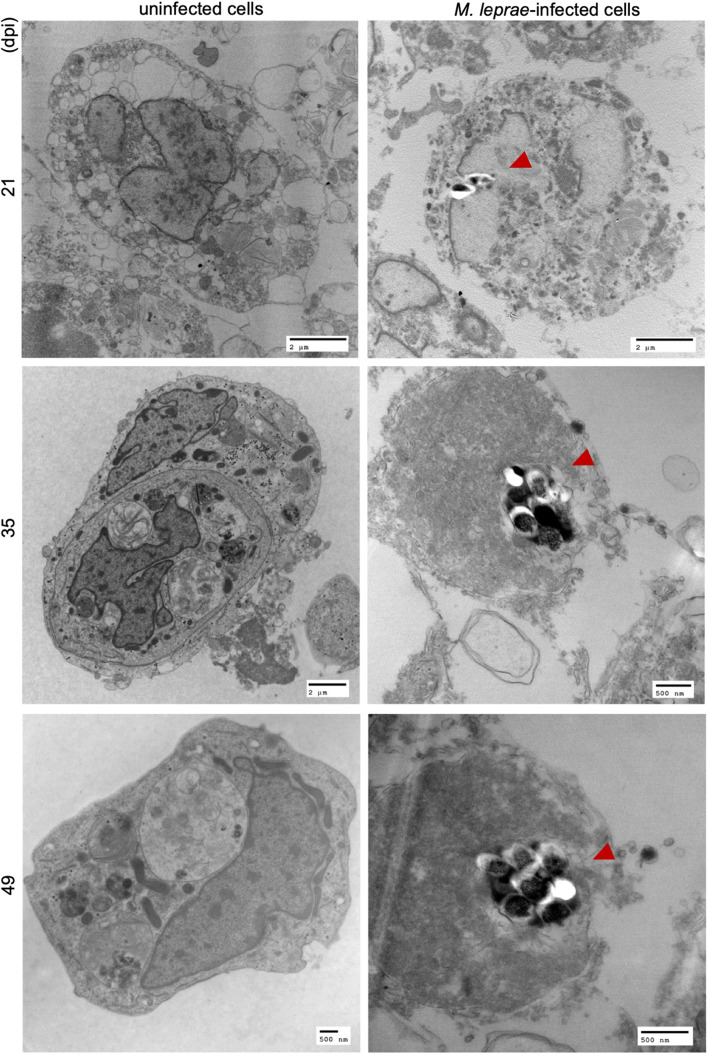
FIGURE 3.
TEM imaging on M. leprae Thai-53 strain infected in tick-derived cell lines. ISE6 cells on coverslips were infected with M. leprae Thai-53 strain. Infected and uninfected samples at 21, 35, and 49 dpi were prepared for TEM imaging. Images were captured using TEM microscope and bacilli were indicated by red arrowheads.

