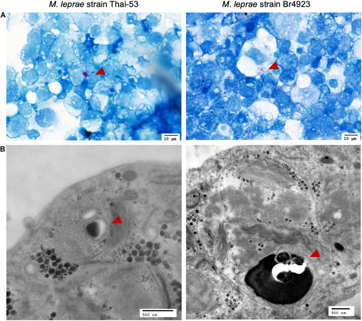
FIGURE 5.
Visualization of M. leprae-infected tick cells by light microscopy and TEM. ISE6 cells infected with M. leprae Thai-53 and Br4923 strains for over 700 days were visualized but light microscopy at magnification 60× after (A) Fite’s acid fast staining and (B) by TEM. Bacilli are indicated by red arrowheads.

