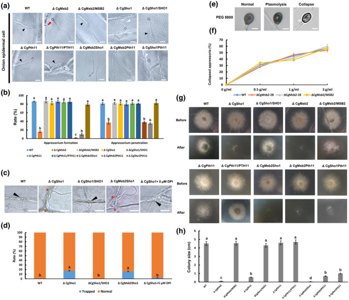
FIGURE 2.

Appressorium formation, penetration, and cellophane membrane penetration assays. (a) Equal volumes (30 μl) of conidial suspension (2 × 10⁴/ml) of each strain were inoculated on onion epidermal cells; the images were acquired at 12 hr postinoculation (hpi). A black triangle indicates an infection hypha. A red arrow indicates an appressorium without penetration. A white triangle indicates a short infection hypha. Bars = 10 µm. (b) Bar chart showing the rate of appressorium formation and penetration on onion epidermal cells at 12 hpi. At least 200 germinated conidia or appressoria from each strain were measured to calculate the appressorium formation or penetration rate, respectively. Error bars represent the standard deviations based on three independent replicates. The values indicated by different letters are significantly different within the same treatment at p < .05, as determined using Tukey's post hoc test. (c) Infection hyphae of wildtype (WT), ΔCgSho1, ΔCgSho1/SHO1, ΔCgMsb2Sho1, and ΔCgSho1 supplied with 5 μM diphenyleneiodonium chloride in onion epidermal cells at 24 hpi. A black triangle indicates an infection hypha breaking through the cell wall of adjacent cells. A red asterisk indicates a trapped infection hypha. Bars = 25 µm. (d) The rate of normal or trapped infection hyphae of each strain at 24 hpi. The values indicated by different letters are significantly different at p < .05, as determined using Tukey's post hoc test. (e) Conidial suspensions (2 × 10⁴/ml) from WT, ΔCgMsb2‐28, ΔCgMsb2‐33, and ΔCgMsb2/ MSB2 were inoculated on the hydrophobic side of a GelBond membrane. At 12 hpi, the water drop was removed by 30 µl of 0.5, 1, and 3 g/ml polyethylene glycol (PEG) 8000. After 5 min of treatment, the collapse rate of appressoria was recorded. There are three types of appressoria: normal, plasmolytic, and collapsed appressoria. A white triangle indicates a collapsed appressorium (longitudinal cavity). Bar = 2 μm. (f) Line chart showing the rate of collapsed appressoria under the treatment of different concentrations of PEG 8000. This experiment was repeated three times. At least 200 melanized appressoria from each strain were measured to calculate the proportion of collapsed appressoria. (g) The strains were grown on cellophane membranes overlaid on potato dextrose agar for 3 days at 25 °C (Before). The cellophane membrane was removed, and the resulting plates were incubated at 25 °C for an additional 2 days (After). (h) Bar chart showing the colony size of each strain at 2 days after removal of the cellophane membrane. Error bars represent the standard deviations based on three independent replicates. The values indicated by different letters are significantly different at p < .05, as determined using Tukey's post hoc test
