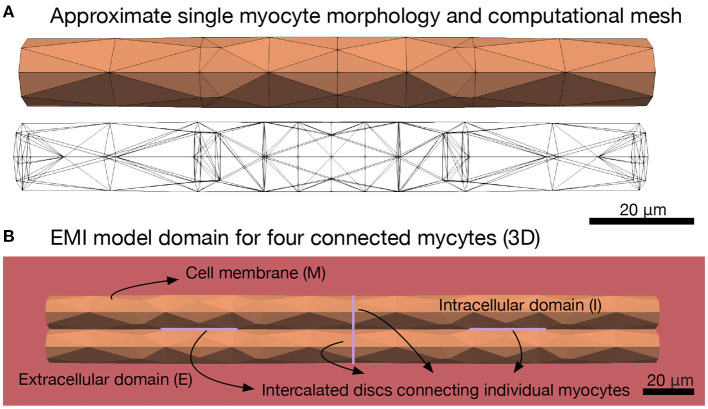
Figure 1.
Illustration of the EMI model domain. (A) Shows the approximate cylindrical geometry and associated mesh of a single myocyte of length 120 μm and diameter ranging from 13 to 14 μm (Nygren et al., 1998). (B) Shows an illustration of the different components of the EMI model domain for an example collection of four connected myocytes. The domain consists of a number of myocytes surrounded by an extracellular space. The cell membrane is defined at the interface between the intracellular and extracellular spaces and intercalated discs with gap junctions are defined at the interface between adjacent myocytes. All computations presented here are in 3D.

