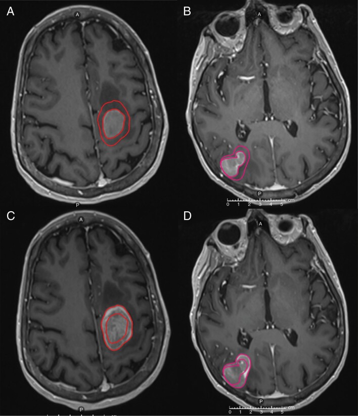
Figure 1.
Axial T1-weighted post-contrast images of a patient with BRAF-mutated metastatic melanoma with brain metastases in the left frontal (A) and right parietal lobes (B) visualized on the diagnostic MRI (MRI-1). Comparison with the treatment planning MRI (MRI-2) performed at SRS 8 days later demonstrated significant interval growth with comparative contours displayed (C, D). Abbreviations: MRI, magnetic resonance imaging; SRS, stereotactic radiosurgery.

