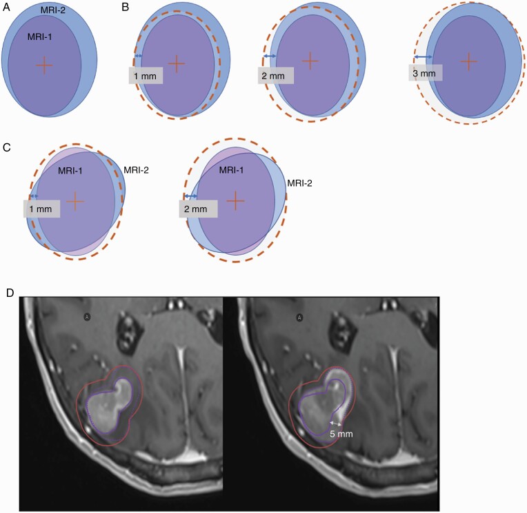
Figure 2.
Schematic 2D illustration of the systematic approach to evaluating tumor dynamics used in this study. An example GTV volume on MRI-1 is shown in purple and in blue to represent the GTV on MRI-2 (A). In the first scenario, there is a tumor volume enlargement observed on comparison on MRI-2 to MRI-1. Consequently, a uniform expansion with 1-, 2-, and 3-mm margin is implemented to ensure target volume coverage. In this example, a 3 mm covered the GTV on MRI-2 (B). In a second scenario, the tumor has not changed in size between the MRIs but has changed in shape, also resulting in undercoverage of the target volume. An expansion margin of 2 mm is needed to ensure target volume coverage (C). Actual case example of a treatment planning MRI for a right parietal brain metastasis treated with SRS. Comparison of initial tumor size required a 5-mm margin to adequately cover the disease extent at the time of treatment (D). Abbreviations: GTV, gross tumor volume; MRI, magnetic resonance imaging; SRS, stereotactic radiosurgery.

