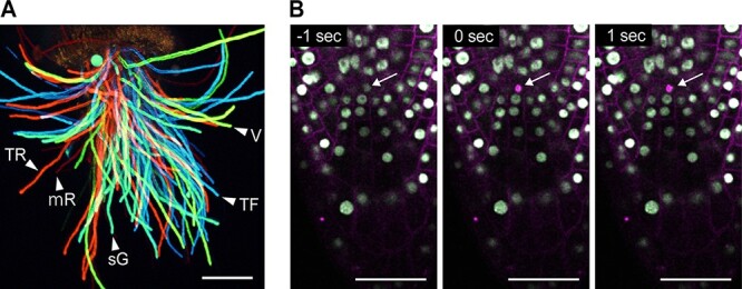
Fig. 2.

Simultaneous multicolor imaging and laser ablation by two-photon excitation. (A) Simultaneous multicolor two-photon imaging of Arabidopsis thaliana pollen tubes. Pollen tubes emerging from the end of cut style were observed 5 h after pollination. Each pollen tube expresses one of the following five fluorescent proteins—mTFP1 (TF), sGFP (sG), Venus (V), TagRFP (TR) or mRFP (mR). Emitted fluorescence signals were detected using a 32-channel PMT array detector ranging from 463.9 to 649.2 nm at 6.0 nm intervals. Maximum-intensity projections of Z-stack images at 0–126 μm depth were captured using 22 z-planes with 6 μm intervals after excitation at 980 nm. Spectrum analysis and adding color were also processed by NIS-Elements (Nikon). The microscope is A1R MP (Nikon). Imaging system and detail of marker lines have been described in a previous study (Mizuta et al. 2015). Scale bars, 100 μm. (B) Laser ablation of a root stem cell as a single cell level on the Arabidopsis thaliana root tips. Two-photon imaging of the root tip of Arabidopsis thaliana expressing RPS5Apro::H2B-sGFP and RRPS5Apro::tdTomato-LTI6b is shown. Excitation and irradiation wavelength used was 980 nm. Time indicates the elapsed time from the start of laser irradiation. Two-photon laser was used to irradiate a root stem cell at the single cell level (arrow) for 0.5 s. Scale bars, (A) 100 μm and (B) 50 μm.
