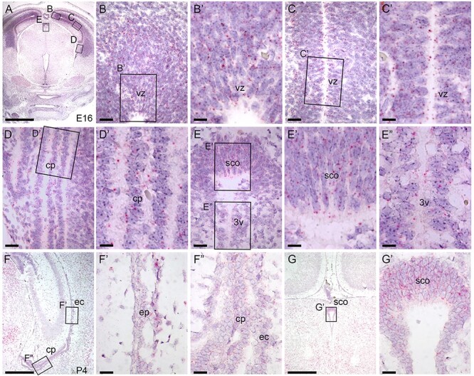
Fig. 2.

B3glct is expressed ubiquitously in brain. RNAscope® analysis of B3glct mRNA expression. Localization of B3glct mRNA appears as red dots overlying cells counterstained with hematoxylin (purple). (A–E″) At E 16.5, B3glct transcripts were localized in the ventricular zone (vz) of the cortex (B–C′), choroid plexus (cp) (D, D′), subcommissural organ (sco) (E, E′) and ependyma of third ventricle (3v) (E″). (F–G′) At P4, B3glct mRNA was localized to ependymal cells (ec) of the lateral ventricle (F′), choroid plexus (cp) (F″) and subcommissural organ (sco) (G, G′). Rectangles indicate the magnified regions of brain sections. Scale bars: panel A, 1 mm; panel F, G, 500 μm; panels B, C, D, E, F′, F″ and G′, 50 μm; and panels B′, C′, D′, E′ and E″, 20 μm.
