Fig. 5.

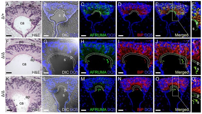
AFRUMA is secreted in B3glct mutant subcommissural organ. (A–O′) Analysis of SSPO trafficking in subcommissural organs from P21 wild-type (Δ/+) (A–E′) and B3glct knockout (Δ/Δ) with mild ventriculomegaly (F–J′) or severe hydrocephalus (K–O′). (A, F, K) Hematoxylin and eosin (H&E)-stained subcommissural organ sections. pc, posterior commissure; bs, basal side; ap, apical side; and ca, central aqueduct. (B, G, L) DRAQ5 (DQ5) staining of subcommissural organ sections merged with differential interference contrast (DIC) image to indicate boundaries of intracellular (i), presecretory (p) and secreted (s) regions of the subcommissural organ. (C–E′, H–J′, and M–O′) Maximum projection images of immunostained subcommissural organ sections stained with AFRUMA (green) and DRAQ5 (blue) (C, H, M) or anti-BiP (red) and DRAQ5 (blue) (D, I, N). Merged channels are shown in (E–E′, J–J′, and O–O′). Rectangles in (E, J, O) indicate regions digitally expanded to the right (E′, J′, O′). Dotted curves demarcate intracellular, presecretory and secreted regions in subcommissural organs. See Figure 6 for quantification of immunofluorescence signals and colocalization of signals. Scale bars: panels A, F, K, 50 μm, and panels B–E, G–J, L–O, 20 μM.
