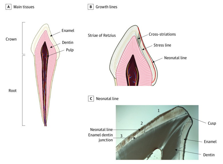
Figure 1. Primary Tooth Development and the Neonatal Line.
A, Primary teeth are composed of 3 main tissues: enamel, dentin, and pulp. Enamel is formed by cells called ameloblasts, which operate in a circadianlike process to lay down the enamel matrix in incremental layers; this incremental growth process is permanently recorded in enamel and dentin through a series of growth lines, which can be observed in longitudinal cross-sections of the tooth using light microscopy.27,28 B, Human teeth show daily growth lines, called cross-striations, which appear between longer period growth lines27 called striae of Retzius28 that correspond to roughly weekly growth. When an insult or disruption occurs during enamel or dentin formation, the growth mark may appear wider or darker; these more pronounced growth marks are referred to stress lines29,30 (or accentuated lines). C, The neonatal line is 1 of the most prominent stress lines, present in approximately 90% of primary teeth and 10% of permanent first molars.31 In this study, the neonatal line width was measured 3 times at 3 locations along the enamel prism: (1) the cuspal third or third closest to the enamel surface (referred to as the cusp), (2) the middle third, and (3) the third closest to the enamel-dentin junction, the point where the enamel and dentin meet.

