Fig. 1.

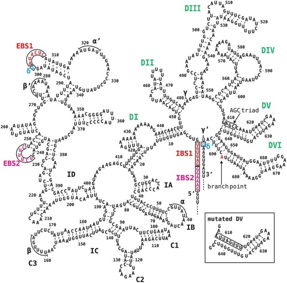
Model of the tobacco atpF intron secondary structure. Domains I–VI are labeled in bold uppercase letters (green). EBSs and IBSs (red, boxed) and δ and δ′ (blue, boxed) are presented. Additional possible interactions (α–α′, β–β′ and γ–γ′) are related to intron compaction. The conserved AGC triad in DV is boxed. The branch point A (red) in DVI is indicated. The inset presents the mutated DV (mDV). The intron and exon sequences are from accession Z00044.2.
