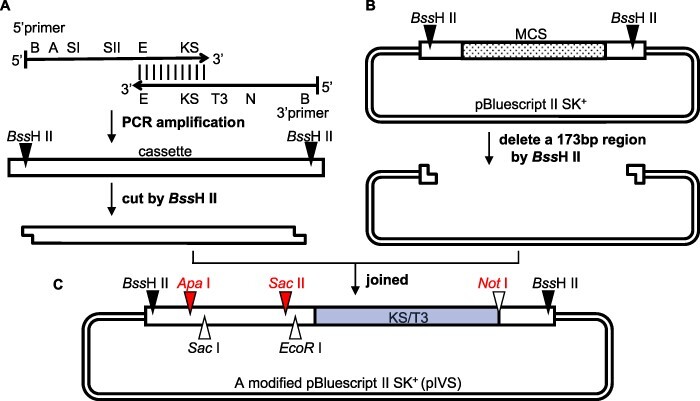
Fig. 2.

Construction of the pIVS vector for preparing pre-mRNAs. (A) A 95-bp cassette was prepared by a PCR amplification involving the 5′ primer containing the BssHII (B) ApaI (A), SacI (SI), SacII (SII) and EcoRI (E) sites and a KS sequence (KS) as well as the 3′ primer with the (E) KS/T3 sequence (KS/T3), NotI (N) and BssHII (B) sites. The KS/T3 sequence consists of a 17-bp KS primer-binding site and a 20-bp T3 promoter from pBluescript II SK + . The primers are complementary between E and KS, and their full sequences are provided in Supplementary Fig. S2. (B) A 173-bp region [box, including the T7 promoter, the multiple cloning sites (MCS, dots), KS/T3] in pBluescript II SK+ was deleted following digestion with BssHII. (C) Schematic of the pIVS vector. The SacI site (triangle) is an alternative site for inserting specific gene fragments (rpl16, rps16, rpoC1, atpF and petB, which have a 5′ UTR with a single SacI site). The NotI (red) is used to linearize the plasmid for transcription, whereas EcoRI is used to digest the templates.
