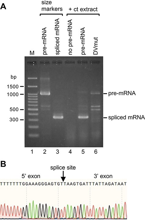
Fig. 5.

Detection of spliced atpF mRNA products after the in vitro splicing reactions. (A) Gel patterns of spliced mRNA products after the 2-h incubation at 28°C. The bands represent the double-stranded DNA produced from the respective mRNAs by RT-PCR amplification (see Fig. 3F). The pre-mRNA (approximately 1,040 nt) and the synthesized spliced mRNA (approximately 340 nt) were used as size markers (lanes 2 and 3). The reaction without the pre-mRNA (lane 4). The spliced mRNA after the reaction (lane 5). No splicing for the pre-mRNA with a mutated Domain V (DVmut, lane 6). (B) Sequencing of the DNA reverse-transcribed from the spliced mRNA (lane 5). A portion of the sequence is presented. The splice site is indicated with an arrow.
