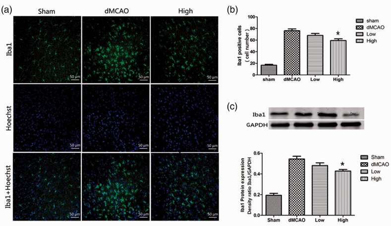
Figure 3.
Artesunate suppressed microglial activation induced by cerebral ischemia. (a) Brain sections were double‐stained with anti-Iba-1 (green) and Hoechst (blue) to label microglia in the cerebral cortex in different groups at 72 hours after distal middle cerebral artery occlusion (dMCAO). (b) Quantification of Iba1-immunopositive cells. *P < 0.05 vs the dMCAO group. (c) Western blotting analysis and quantification of the Iba1 protein level in the cerebral cortex in different groups (*P < 0.05 vs the dMCAO group).

