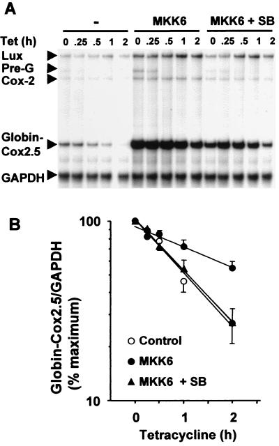
FIG. 2.
The Cox-2 3′ UTR mediates regulation of mRNA stability by the p38 pathway. HeLa-TO cells were transfected with 200 ng of pGL3c, 20 ng of pTetBBB-Cox2.5, and 100 ng of MKK6 expression vector or empty vector control (pcDNA3). After 24 h vehicle control (dimethyl sulfoxide) or SB203580 (1 μM) was added. After a further 30 min, tetracycline was added to a final concentration of 100 ng/ml. Cells were harvested at the time intervals shown, and ribonuclease protection assays were performed to quantify luciferase, Cox-2, β-globin–Cox2.5, and GAPDH mRNAs. (A) A representative experiment. Ribonuclease protected luciferase (Lux), Cox-2, GAPDH, β-globin–Cox2.5 and pre-β-globin (Pre-G) fragments are indicated. (B) Graphical representation of means ± standard deviations (error bars) of seven independent experiments. β-globin/GAPDH ratios were plotted as percentage of the maximum value at the time of tetracycline addition.

