Fig. 1.
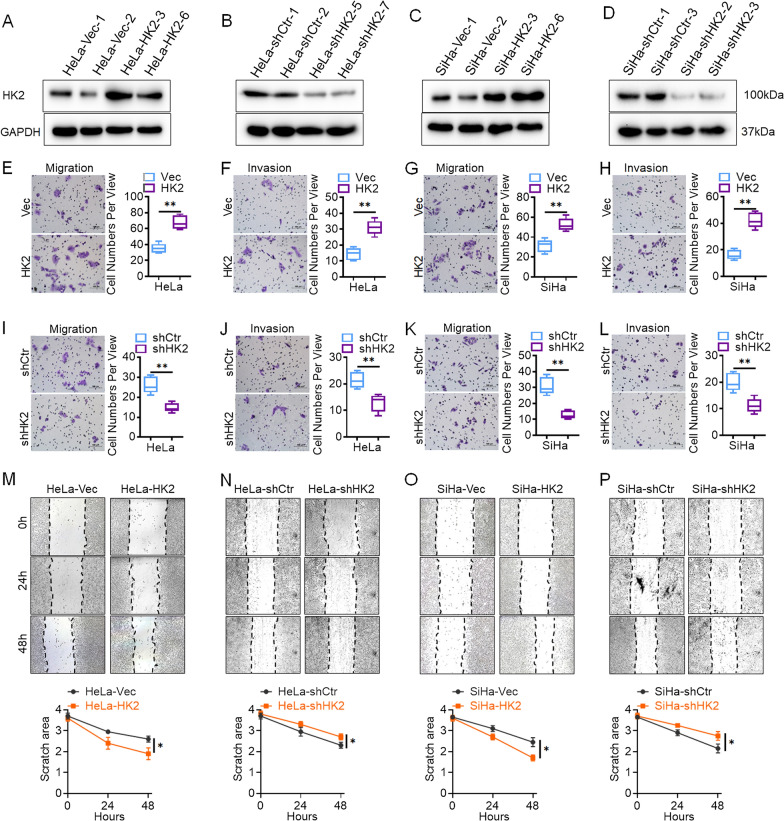
HK2 enhances the migration and invasion ability of SiHa and HeLa cells in vitro. Stably transfected cell lines were identified by western blotting: A HeLa-Vec and HeLa-HK2 cells; B HeLa-shControl and HeLa-shHK2 cells. C SiHa-Vec and SiHa-HK2 cells; D SiHa-shControl and SiHa-shHK2 cells. The migratory capacities were analyzed by the transwell assay, and the number of migratory cells is shown (scale bar, 100 μm). E HeLa-Vec and HeLa-HK2 cells G SiHa-Vec and SiHa-HK2 cells. I HeLa-shCtr and HeLa-shHK2 cells K SiHa-shCtr and SiHa-shHK2 cells. The invasive capacities were analyzed by the transwell assay, and the number of migratory cells is shown (scale bar, 100 μm). F HeLa-Vec and HeLa-HK2 cells H SiHa-Vec and SiHa-HK2 cells. J HeLa-shCtr and HeLa-shHK2 cells L SiHa-shCtr and SiHa-shHK2 cells. The migratory potential was analyzed by wound-healing assays performed for 0, 24, and 48 h. M HeLa-Vec and HeLa-HK2 cells N HeLa-shCtr and HeLa-shHK2 cells O SiHa-Vec and SiHa-HK2 cells. P SiHa-shCtr and SiHa-shHK2 cells (scale bar, 200 μm). The data are shown as the mean ± SD of three independent experiments. *p < 0.05, **p < 0.01 vs. control using one-way ANOVA

