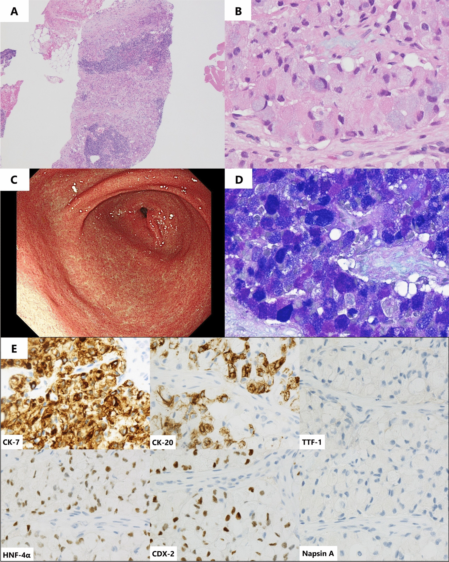
Fig. 3.

Pathological findings of right cervical lymph node biopsy. A Pathological findings of right cervical lymph node biopsy with hematoxylin and eosin staining indicate tumor cells in the specimen (×40) as well as B tumor cells with a signet ring cell feature (×400). C Upper endoscopy shows no abnormal findings in the pyloric region of the stomach. D Lymph node biopsy with Alcian blue-periodic acid-Schiff staining shows mucin-abundant tumor cells (×400). E Immunohistochemical staining shows positive cytokeratin (CK)-7, positive CK-20, negative thyroid transcription factor-1 (TTF-1), positive hepatocyte nuclear factor-4α (HNF-4α), positive caudal-type homeobox-2 (CDX-2), and negative napsin A (×400)
