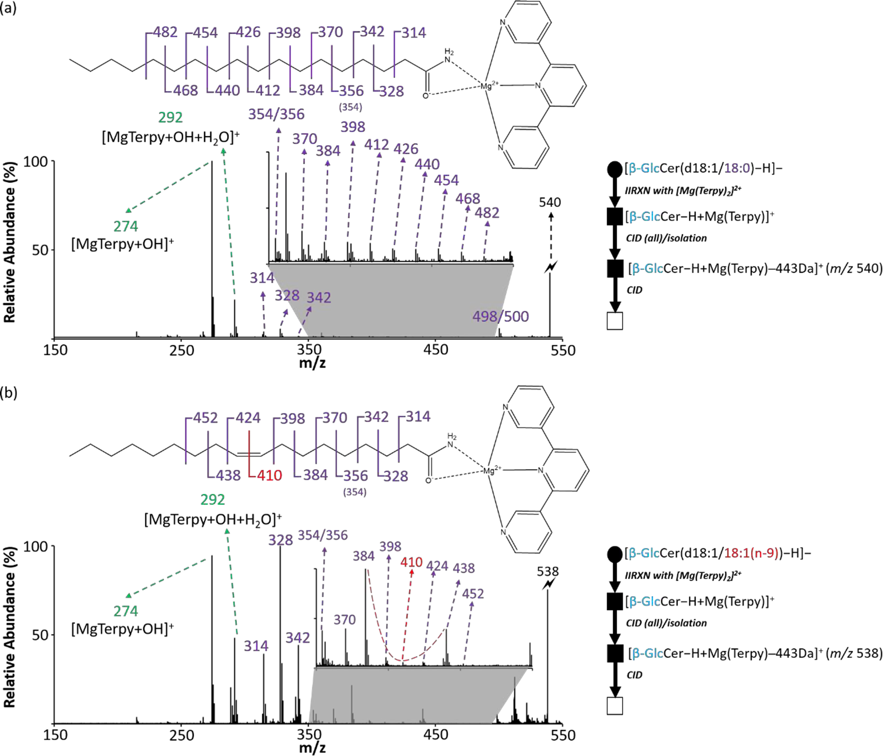
Figure 5.

The identification of double bond position from the monounsaturated fatty acyl side chain on cerebrosides. (a) The CID spectrum of 443 Da loss ion from [β-GlcCer(d18:1/18:0)−H + MgTerpy]+. (b) The CID spectrum of 443 Da loss ion from [β-GlcCer(d18:1/18:1 (n-9))−H + MgTerpy]+. The inserts are the zoom-in spectra of m/z region ranged from 350 to 500. The red dashed line signifies the special spectral gap pointing the double bond position. The symbols represent as same as those in Figure 2.
