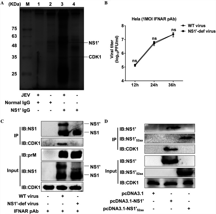
FIG 1.
Identification of interaction between JEV NS1′ and host CDK1 protein. (A) HeLa cells were infected with WT JEV at an MOI of 1.0 for 36 h followed by the co-IP analysis of cell lysates with JEV NS1′ antibodies. The purified proteins were visualized by silver staining. The protein bands in lane 3 which are different from those in lane 4 were excised and analyzed using the LC-MS/MS. The position and names of proteins identified by MS are indicated. (B) HeLa cells treated with IFNAR polyclonal antibodies (pAb) were mock-infected or infected with WT virus or NS1′-def virus at an MOI of 1.0, and the virus titers in the culture supernatants were measured by plaque assay at 12, 24, and 36 hpi. (C) HeLa cells were treated with IFNAR pAb and then infected with WT virus or NS1′-def virus at an MOI of 1.0 for 36 h, followed by co-IP analysis of cell lysates with JEV NS1 antibodies. Cellular proteins coimmunoprecipitated by respective antibodies were analyzed by Western blotting. (D) HeLa cells were transfected with pcDNA3.1 plasmids expressing the NS1′, ΔNS1′52aa, or empty vector for 36 h, followed by co-IP analysis of cell lysates with JEV ΔNS1′52aa antibodies. Cellular proteins coimmunoprecipitated by respective antibodies were analyzed by Western blotting. All data in panels B to D were pooled from three independent experiments. ns, nonsignificant.

