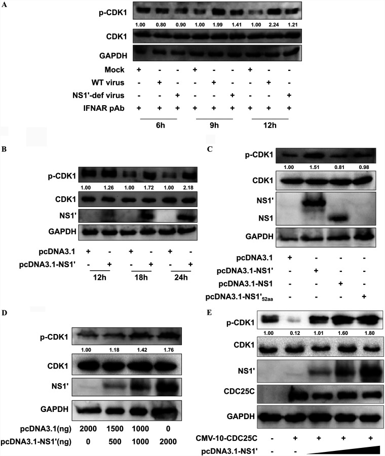
FIG 2.
JEV NS1′ enhances the CDK1 phosphorylation by reducing the CDC25C function. (A) HeLa cells treated with IFNAR pAb were mock-infected or infected with WT virus or NS1′-def virus at an MOI of 1.0, and then the cells were harvested at 6, 9, and 12 hpi. Whole-cell protein lysates were separated by SDS-PAGE and examined by Western blotting using indicated antibodies. (B) HeLa cells were transfected with pcDNA3.1-NS1′ or empty vector for 12, 18, and 24 h, and cell lysates were subjected to Western blotting using indicated antibodies. (C) HeLa cells were transfected with pcDNA3.1 plasmids encoding the JEV NS1, NS1′, or ΔNS1′52aa, or empty vector for a period of 18 h. Total cell lysates were collected for analysis by Western blotting with indicated antibodies. (D) HeLa cells were cotransfected with empty vector and an increasing amount (0, 500, 1,000, or 2,000 ng) of pcDNA3.1-NS1′. The expression of p-CDK1, CDK1, and JEV NS1′ protein was detected by Western blotting with indicated antibodies. (E) HeLa cells were cotransfected with p3×FLAG-CMV-10-CDC25C and different dosages of pcDNA3.1-NS1′. Cells were harvested at 24 h posttransfection and were subjected to Western blotting with indicated antibodies. Protein levels of p-CDK1 were quantified by immunoblot scanning using image J software and normalized to the amount of GAPDH.

