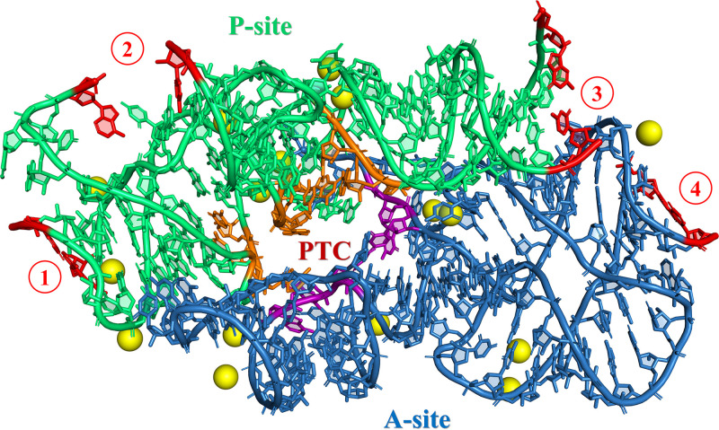
FIG 3.
Three-dimensional representation of the pseudosymmetrical region (SymR; Agmon et al. [250]) extracted from the Thermus thermophilus crystallographic structure (NCBI ID 4WPO) using PyMOL (The PyMOL Molecular Graphics System, version 2.0; Schrödinger, LLC). The residues from the P-site and the A-site are colored green and blue, respectively. Residues delineating the entrance to the PTC pore are colored orange (P-site) and purple (A-site), as defined by Fox et al. (22). Sixteen conserved magnesium ions that are around the contact area of the SymR residues are shown as yellow spheres, as described by Rivas and Fox (68). Regions where the addition of fragments is required for the ligation of smaller fragments are highlighted by red numbers within red circles at the end of each helix, three at the P-site and one at the A-site. Also, the last residues of each ligation point are colored red. A secondary structure depiction of these elements is presented in Fig. 4.

