Figure 6.
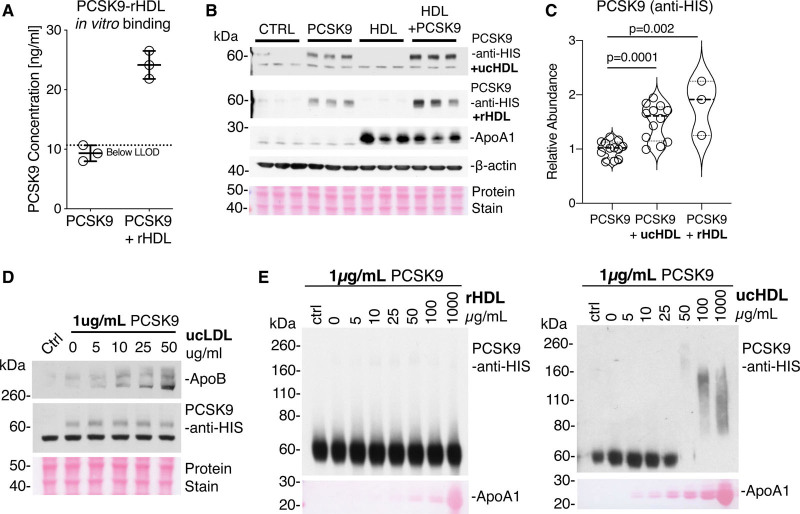
HDL (high-density lipoprotein) potentiates PCSK9 (proprotein convertase subtilisin/kexin type 9) uptake and multimerization. A, PCSK9 and rHDL (reconstituted HDL) were coincubated before HDL immuno-isolation to demonstrate the interaction between rHDL and PCSK9. PCSK9 alone was run through the HDL immunoisolation column as a negative control. B, HepG2 cells were treated with HIS (polyhistidine)-tagged PCSK9 (5 µg/mL), rHDL, or ucHDL (ultracentrifuge-isolated HDL; 25 µg/mL) or a combination of rHDL or ucHDL and HIS-tagged PCSK9 for 6 h before immunoblot analysis for PCSK9 cellular uptake. C, Densitometry analysis of 3 independent replicates. Significance was determined using an independent t test. D, HepG2 cells were treated with HIS-tagged PCSK9 (1 µg/mL), in the presence of increasing amounts of ucLDL (ultracentrifuge-isolated low-density lipoprotein; 0–50 µg/mL). The control lane (Ctrl) represents PCSK9 only. E, Recombinant HIS-tagged PCSK9 at a concentration of 1 µg/mL was incubated in the presence of an increasing concentration of rHDL or ucHDL. Immunoblot analysis was then conducted, with equal amounts of PCSK9 loading for each sample. The control lane represent PCSK9 alone incubated at 4°C. Total protein stain is used to visualize apoA1. LLOD indicates lower limit of detection.

