FIG 3.
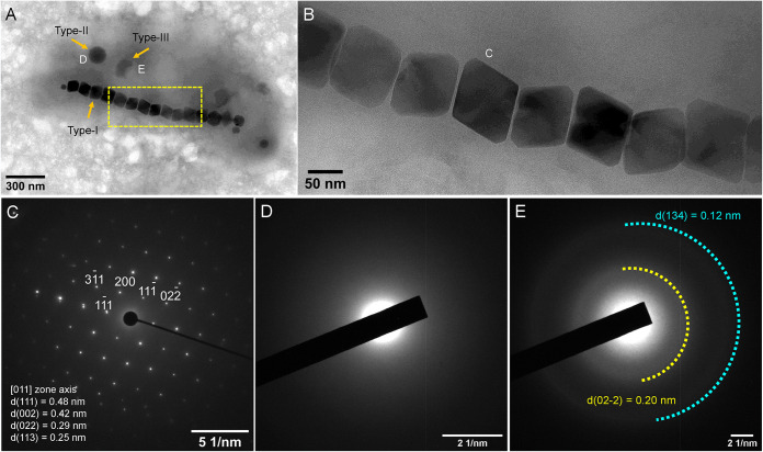
Morphological and structural features of intracellular inclusions within an XQGS-1 cell. (A) TEM image of an XQGS-1 cell that contains three electron-dense particle types. (B) Close-up view of an XQGS-1 magnetosome chain indicated by the yellow dashed box in panel A. (C to E) Selected-area electron diffraction (SAED) patterns recorded from an individual magnetite particle (C) (labeled “C” in panel B), a type II granule (D) (labeled “D” in panel A), and a type III particle (E) (labeled “E” in panel A). SAED analyses indicate that the magnetic particles are well-crystallized single crystals with d-spacings of 0.48 nm and 0.42 nm, which correspond to the interplanar spacings (d) of the {111}, {002}, {022}, and {113} planes of magnetite, respectively. In contrast, the SAED pattern in panel D for type II granules has no obvious diffraction rings or spots, which is typical of amorphous phases, while the SAED pattern in panel E has two weak, smeared diffraction rings, which indicates that type III inclusions are weakly crystalline.

