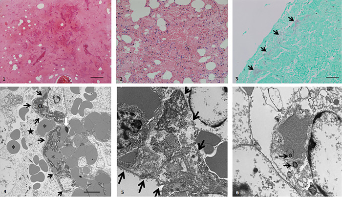
FIG 1.
Light and electron microscopy of lung tissue from case 1 of hemorrhagic pneumonia due to Escherichia coli. (Panel 1) (Hematoxylin and eosin [H&E] stain) Near-total effacement of normal architecture by hemorrhage, edema, fibrin, and necrotic debris. Bar, 200 μm. (Panel 2) (H&E stain) Alveolar walls are effaced. Alveolar spaces are filled with hemorrhage, edema, fibrin, and pyknotic mononuclear cells. Bar, 100 μm. (Panel 3) (Gram stain) Gram-negative bacilli (arrows) aggregate within alveolar spaces abutting the pleura. Bar, 50 μm. (Panel 4) (Transmission electron micrograph [TEM]) Alveolar space (star) contains erythrocytes (asterisks) and alveolar capillaries (black arrows). Capillary lumen (CL) contains fibrin thrombus. White arrow, pyknotic capillary endothelial cell. Bar, 5 μm. (Panel 5) (TEM) Alveolar wall (arrows) is expanded by fibrin strands and cell debris. Bar, 2 μm. (Panel 6) (TEM) Macrophage with bacteria in a phagolysosome (black arrows). Bar, 1 μm.

