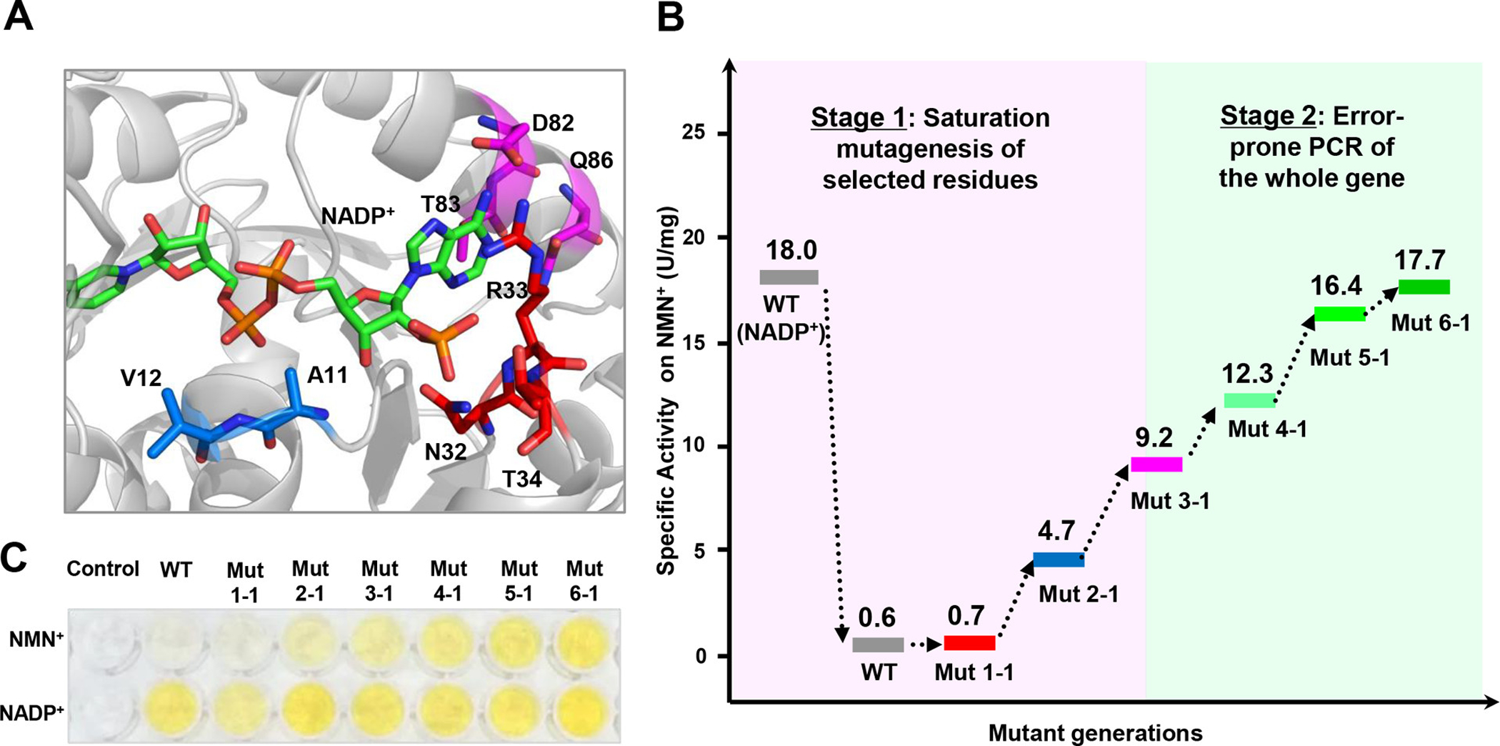
Figure 4.

Directed evolution to improve 6PGDH activity on NMN+. (A) Structural model of wild-type 6PGDH (WT) in complex with NADP+. Residues within <5.5 Å of the 2′ phosphate, pyrophosphate, or adenine moieties of NADP+ are colored red, blue, and magenta, respectively. (B) Evolutionary progress of mutant activities on NMN+. (C) Images of activity measurement of the WT and mutants on NADP+ and NMN+ based on the WST-1/GsDI assay.
