Figure 4.

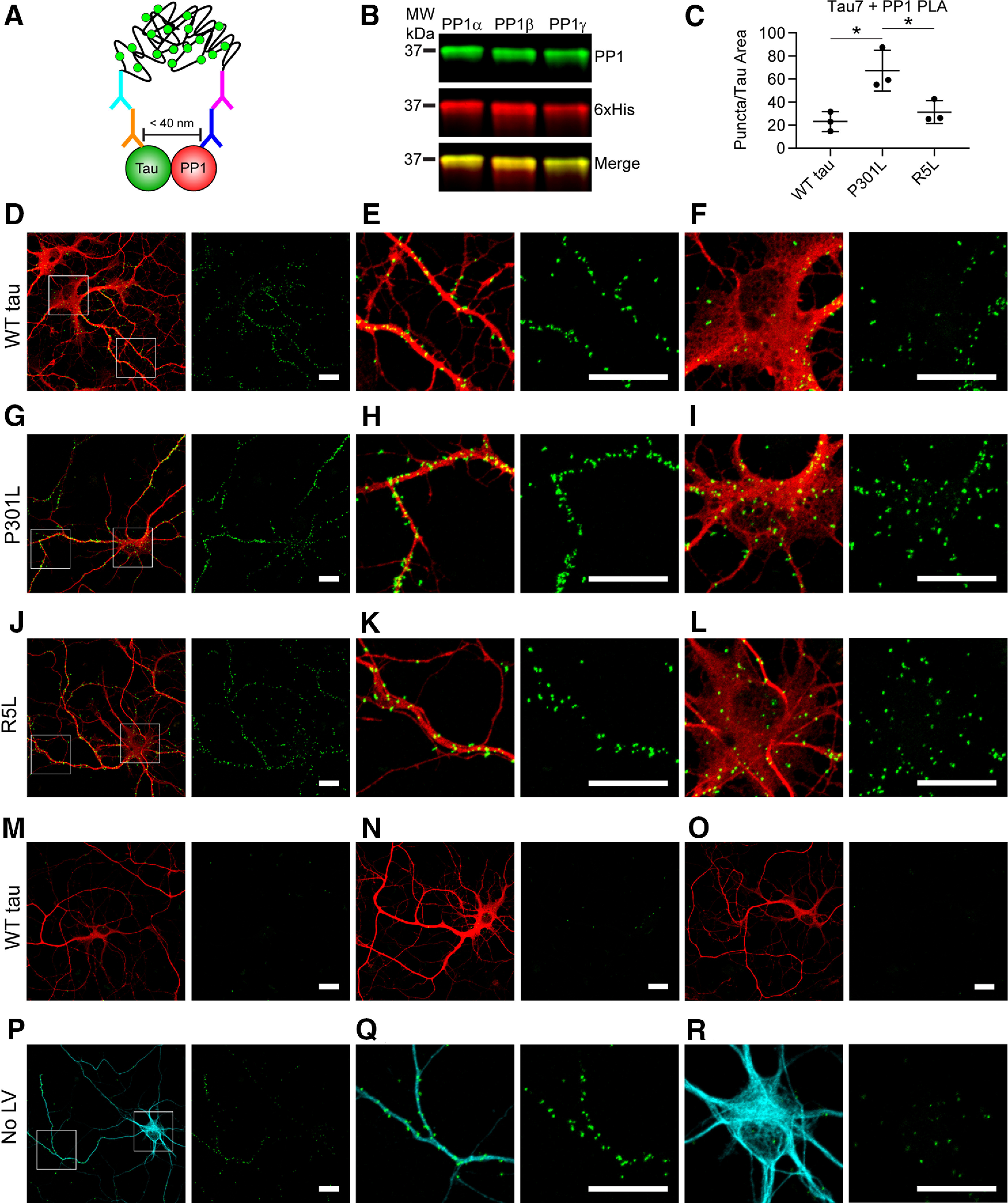
P301L tau increases the association with PP1 in primary hippocampal neurons. A, Lentiviruses were used to express WT, P301L, or R5L tau in cultured primary rat hippocampal neurons beginning at DIV 4 and fixed in 4% paraformaldehyde on DIV 8. The neurons were probed with Tau7 (C-terminal tau epitope) and a PP1 antibody before performing the PLA staining procedure that demonstrates a close association between these proteins (<40 nm) indicated by a green fluorescent punctate signal. Following PLA, the cells underwent immunocytofluorescence staining with a Tau12 (red, human tau-specific N-terminal tau epitope) to identify exogenous tau and Tuj1 (cyan) to identify neurons and neuron processes. B, The PP1 antibody recognizes recombinant PP1α, PP1β, and PP1γ in an immunoblot (green), and a 6× His antibody labels total protein. C, Images of isolated neurons were used to quantify puncta per area of Tau12 staining (i.e., transduced neurons). P301L tau-expressing neurons (67.3 ± 17.7 puncta/nm2) showed significantly greater PLA signal than WT (23.1 ± 8.5 puncta/nm2) and R5L tau-expressing neurons (31.3 ± 9.8 puncta/nm2), complementing results obtained in HEK cell pull down and NanoBRET assays (Figs. 2, 3). Data represent mean ± SD and were analyzed using repeated measures one-way ANOVAs and Tukey's post hoc multiple comparison test, *p ≤ 0.05. D–F, PLA detected associations between WT tau and endogenous rat PP1 (PLA, green puncta; Tau12, red), in both neuronal processes (E) and cell bodies (F). G–I, Neurons expressing P301L tau displayed an enhanced association between tau and PP1 as evidenced by the PLA puncta, which were found in processes (H) and cell bodies of neurons (I). J–L, Neurons expressing R5L tau showed an association between tau and PP1 that was similar to WT tau cells and localized to neuronal processes (K) and cell bodies (L). M–O, Primary antibody delete controls included omission of either Tau7 (M), PP1 (N), or both (O). Primary delete controls showed little to no nonspecific background signal. P–R, PLA in untransduced neurons (No LV) demonstrated an association between endogenous rat tau and PP1, which were evident in neuronal processes (Q) and cell bodies (R) of neurons. Scale bars: 20 μm for all images.
