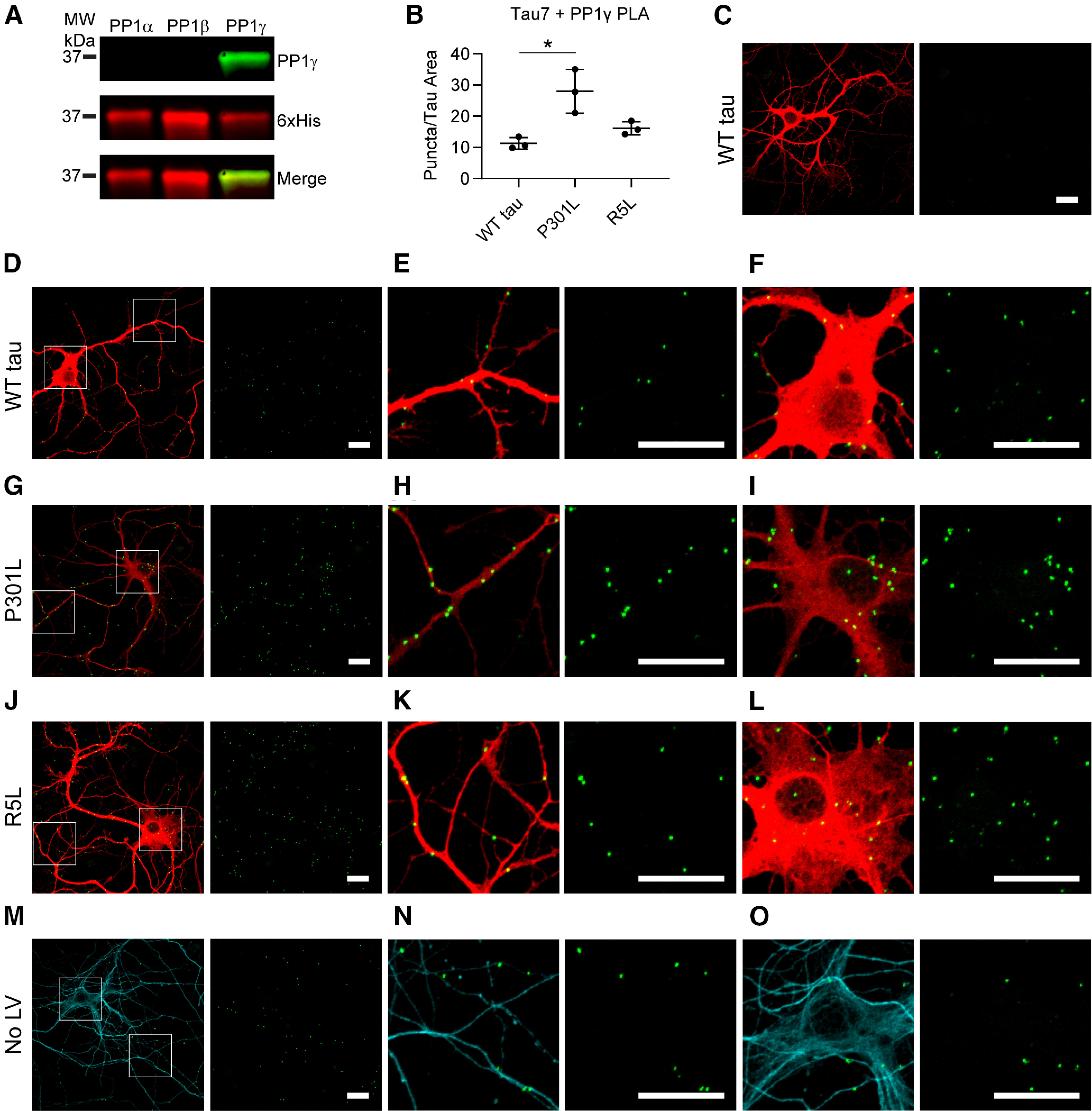
Figure 8.

P301L tau increases the association with PP1γ isoform in primary hippocampal neurons. A, PLA experiments were repeated with the Tau7 antibody and a PP1γ-specific antibody verified with immunoblot. Recombinant PP1 isoforms were probed with a PP1γ antibody (green) and a 6xHis antibody (red). B, PLA puncta were quantified and normalized to area of Tau12 staining (i.e., exogenous tau staining). P301L tau (28.0 ± 7.0 puncta/nm2) expression increased levels of PLA signal compared with WT tau (11.3 ± 1.9 puncta/nm2), whereas R5L (16.2 ± 2.1 puncta/nm2) expression did not significantly change. Data represent mean ± SD of three replicates and were analyzed using a repeated measures one-way ANOVA and Tukey's post hoc multiple comparison test; *p ≤ 0.05. C, A Tau7 primary antibody deletion demonstrated the lack of background signal when PLA is performed with the PP1γ antibody only. D–F, Associations between WT tau and endogenous rat PP1γ (PLA, green puncta; Tau12, red) were identified in the neuronal processes (E) and cell bodies (F) of the neurons. G–I, Lentiviral expression of P301L tau increased PLA puncta, which were found in processes (H) and cell bodies of neurons (I). J–L, PLA in the presence of R5L tau also demonstrated an association between tau and PP1γ, localized to the processes (K) and cell bodies (L) of the neurons. M–O, PLA was performed in untransduced neurons to identify an association between endogenous rat tau and PP1γ, also found in neuronal processes (N) and cell bodies (O). Scale bars: 20 μm for all images.
