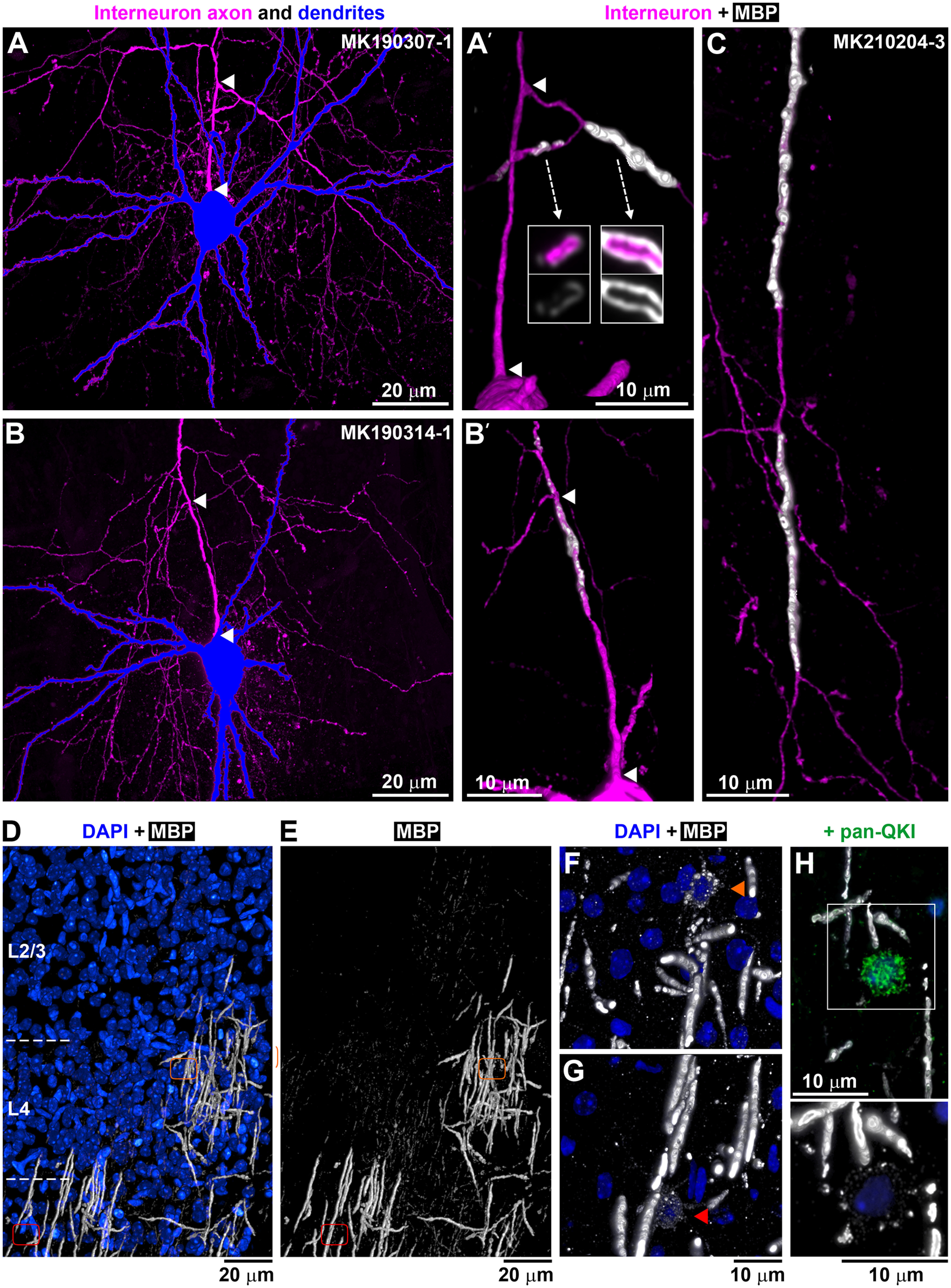
Figure 2.

Axonal branching and myelination at P14. A, B, PV+ interneurons from P14 neuronal pairs, with the soma and dendrites in blue and axon in magenta. MAX projections through all sections from pairs MK190307-1 (775 sections) and MK190314-1 (375 sections). A′, B′, Volume reconstruction of the axon initial segment of the interneurons in A (103 sections) and B, respectively (136 sections). Arrowheads indicate the corresponding beginning and end of the first axonal segment from the soma to the first branching point. A′, There are two tertiary axonal branches that are myelinated; the one going left is incompletely myelinated as seen by the uneven and weak MBP immunolabel. Arrows indicate single sections through the axonal branches. No myelination is observed before the first branching point. B′, There is incomplete myelination before the first branching point as well as after that. No complete myelin was seen in this PV+ interneuron. C, Volume reconstruction of a tertiary axonal branch of the interneuron from pair MK210204-3 (192 sections), showing two completely myelinated internodes (39 and 36 µm long), separated by an unmyelinated axonal stretch (19 µm). D, E, MBP immunostaining of layers 2/3 and 4 at P14 (white; volume reconstruction from 529 sections). Blue represents DAPI-labeled nuclei. Areas with complete, incomplete, and no MBP immunolabel are seen. Orange and red outlines represent the locations of two different myelinating oligodendrocytes shown at higher magnification in F and G. F, G, Myelinating oligodendrocytes characterized by weak MBP immunolabel within their cell bodies and proximal cell processes. Volume reconstruction from 36 sections (F) and 73 sections (G). H, Weakly MBP-immunofluorescent oligodendrocytes (bottom, 13 sections volume reconstruction) colabeled with an antibody against panQKI, an oligodendrocytic marker (top, lower magnification).
