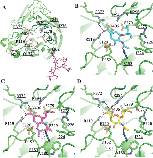
Figure 1.
The structures of N9 neuraminidase (NA) and its complexes (the avian N9 numbering scheme is used). The 10 residues that were substituted in this study are underlined (Table 2). A, Overall structure of the NA monomer, with highly conserved residues of the active site presented as salmon sticks. Residues Q137, T249, and H276 are shown as cyan sticks. Two occupied glycosylation sites are labeled, and glycans are shown as magenta sticks. B, The active site with oseltamivir. C, The active site with zanamivir. D, The active site with sialic (neuraminic) acid. The dashed lines are hydrogen bonds. The structural figure was generated with MacPyMol.

