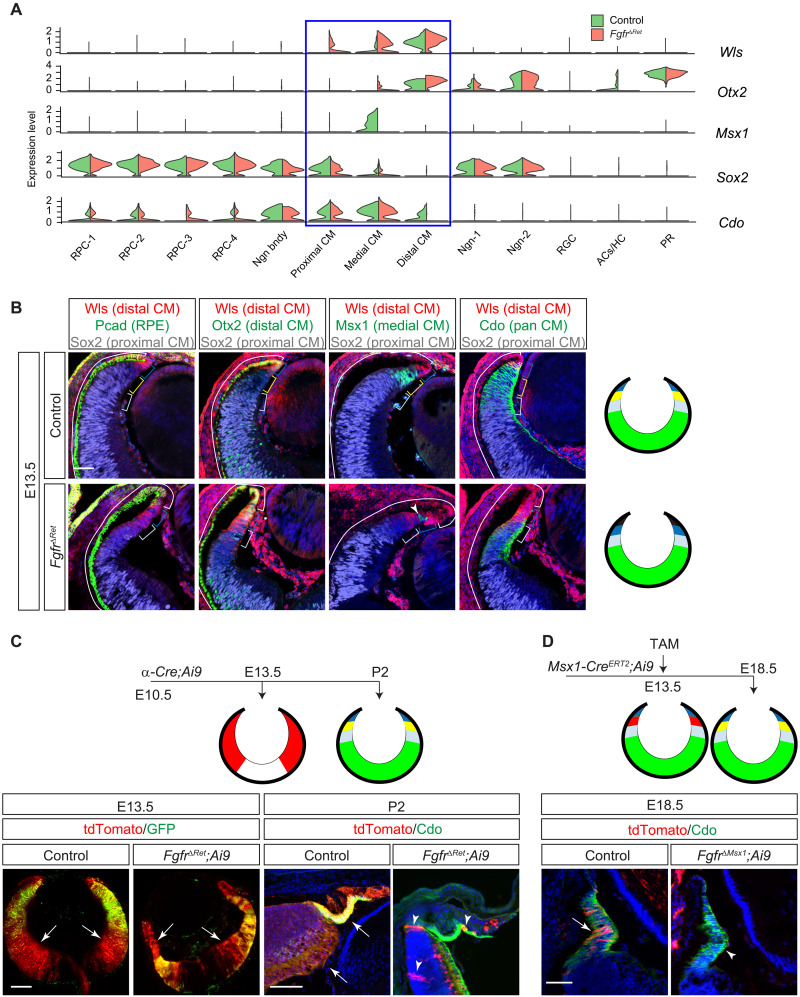
Fig. 3. FGF signaling is required for CM subdivision and survival.
(A) Violin plots showed that the CM can be subdivided by the overlapping expression of Wls, Otx2, Msx1, Sox2, and Cdo, all of which were dysregulated in FgfrΔRet mutants. (B) Immunostaining confirmed that the in silico clustering of CM cells matches the spatial separation of CM subdomains. Aberrant invasion of Pcad, expansion of Wls and Otx2, loss of Msx1, and reduction in Sox2 and Cdo in FgfrΔRet mutants demonstrated CM differentiation defects. Brackets indicate the domain of the RPE and the distal, medial, and proximal CM, corresponding to black, dark blue, yellow, and light blue regions, respectively, in diagrams on the right. The NR is indicated in green. Arrowhead marks residual wild-type cells still expressing Msx1. Scale bar, 50 μm. (C) At E13.5, although Pax6 α-Cre was expressed only in the peripheral retina indicated by its GFP reporter, it has already activated tdTomato expression (arrows) from the Ai9 Cre reporter throughout the retina. At P2, these tdTomato+ progenies remained in control retinae, but only a few (arrowheads) were left in FgfrΔRet mutants. The tdTomato expression is indicated in red in diagrams. Scale bars, 100 μm. (D) CM cells were pulse-labeled by tamoxifen induction of Msx1-CreERT2 at E13.5 and detected at E18.5 by tdTomato expression from the Ai9 Cre reporter. Although these cells remained in the control CM identified by Cdo expression (arrow), they had largely disappeared in the FgfrΔRet mutant (arrowhead), suggesting cell survival defects. Scale bar, 50 μm.

