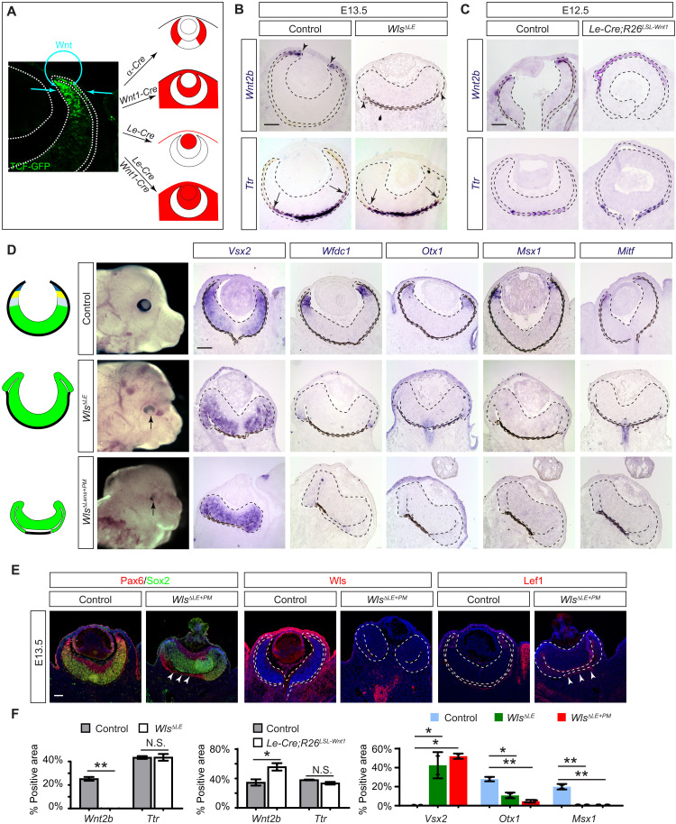
Fig. 6. Paracrine Wnt signaling patterns the CM and the distal RPE.
(A) Potential source of the Wnt signaling gradient revealed by the TCF-GFP reporter may be targeted by α-Cre for retina, Wnt1-Cre for periocular mesenchyme, and Le-Cre for the lens ectoderm (the surface epithelium and the lens). (B) Ablation of Wnt transporter Wls in the lens ectoderm specifically disrupted the proximal RPE differentiation in WlsΔLE mutants as indicated by loss of Wnt2b but not the proximal RPE marker Ttr. (C) Overexpression of Wnt1 in the lens ectoderm also affected the proximal RPE by expanding the domain of Wnt2b expression in Le-Cre;R26LSL-Wnt1 eye cup without affecting Ttr expression. (D) CM was lost in WlsΔLE mutants as shown by the expansion of Vsx2 and the absence of Wfdc1, Otx1, Msx1, and Mitf. The band of the RPE was further diminished in WlsΔLE+ΔPM mutants. (E) WlsΔLE+ΔPM mutant eyes lost Wls and Lef1 in both the lens ectoderm and the periocular mesenchyme, leaving only a small band of Lef1 expression next to the Pax6-expressing RPE (arrowheads). Scale bars, 100 μm. (F) Relative area expressing Wnt2b, Ttr, or Vsx2 was normalized against the entire RPE region, while the Otx1- or Msx1-expressing area was normalized against the NR region. Student’s t test for Wnt2b and Ttr and one-way ANOVA test for Vsx2, Otx1, and Msx1. *P < 0.05 and **P < 0.01; n = 2 for all markers. N.S., not significant.

