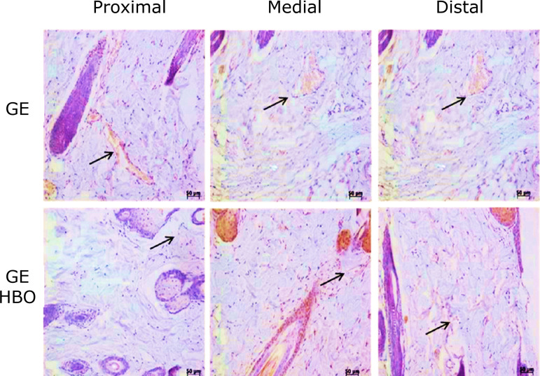
Figure 2. Photomicrographs of histological sections of skin fragments (proximal, medial, and distal regions) from GE and GE/HBO groups, subjected to immunohistochemistry for VEGF-A detection and counterstained with Harris’ hematoxylin. A similar pattern of blood vessels (arrows) immunopositive to VEGF-A were noticed in all analyzed regions. Magnification: x200.
GE: animals submitted to epilation; GE/HBO: animals submitted to epilation subjected to hyperbaric oxygenation; VEGF-A: vascular endothelial growth factor A.

