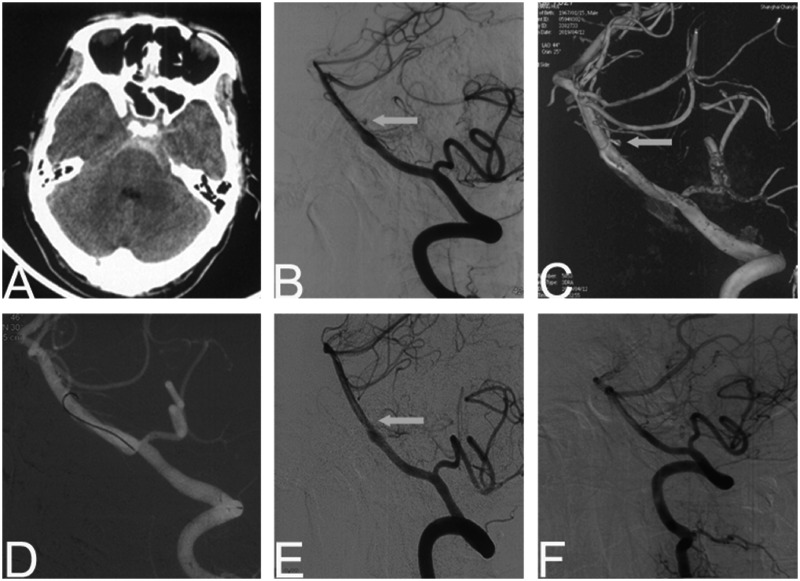
Figure 3.
Patient 3. A 52-year-old man presented with sudden headache. Cranial CT revealing diffuse SAH (a). Cerebral angiography and three-dimensional representation revealing a perforator aneurysm (1.3 mm) near the fenestration of basilar artery (b and c). The electrothrombosis was performed for 1.5 min (d) and the aneurysm disappeared (e). At six-month follow-up, DSA showed no signs of aneurysm recurrence (f).

