
Figure 7. Effects of three peptides derived from AGGF1 functional angiogenic domain on HUVEC adhesion and capillary tube formation.
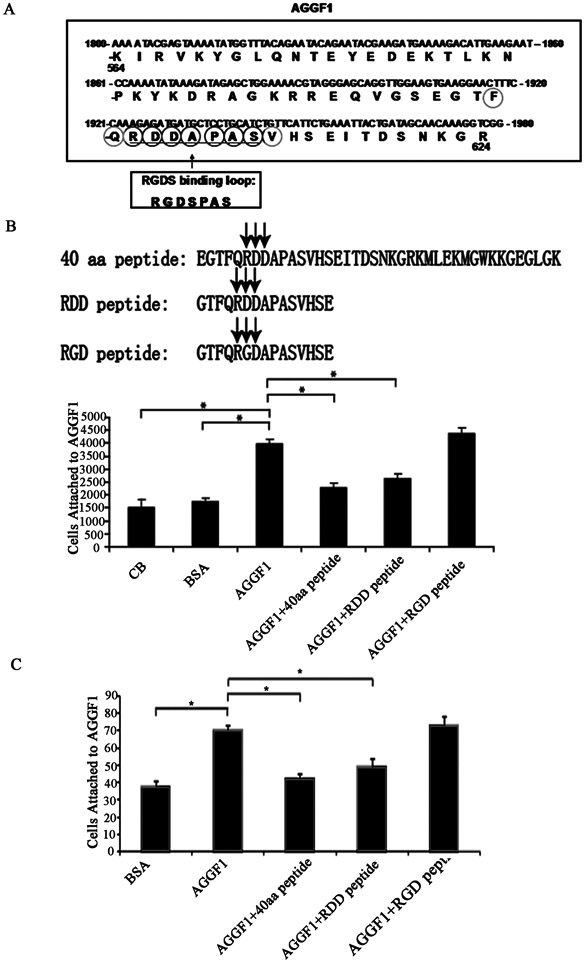
(A) A diagram showing the location and amino acid sequences of the functional Angiogenic Domain of AGGF1. The RDDAPAS motif of AGGF1 is similar, but not identical, to the RGDSPAS integrin binding loop of fibronectin.
(B) HUVEC-AGGF1 adhesion assays with three different AGGF1 peptides. HUVECs were pre-incubated with a peptide (0.12 mM) before being added to the wells coated with AGGF1. Coating buffer (CB) with and without BSA was used as negative controls.
(C) Endothelial tube formation assays with HUVECs treated with the AGGF1 protein in combination with or without three different peptides. HUVECs were pre-incubated with a peptide (0.12 mM) before being added to the wells containing solidified matrigel with WT AGGF1. Matrigel with BSA was used as negative control (left panel).
The data are shown as mean ± SEM. *P≤0.05, n=4/group (one-way ANOVA with Dunnett post hoc tests).