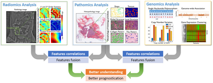
Figure 1.
An overview for the fusion of pathomics, radiomics and genomics analyses. In radiomics analysis, quantitative image features were derived from radiology images, which may include traditional hand-crafted features, e.g., 1st and 2nd order statistics, Laws & Local Binary Patterns, Gradient orientations and Gabor and features that learnt by deep learning model. In pathomics analysis, quantitative image features were derived from histopathological images, which may include hand-crafted features like nuclear shape, texture, global structure, local structure, stroma collagen pattern and TIL patterns and features that learnt by deep learning model. In genomics analysis, single nucleotide polymorphism (SNP), copy number variation (CNV), genome structure data and gene expression data [e.g., ribonucleic acid (RNA)-seq data] were analyzed. In the context of prognosis, features/signatures that associated with patient outcomes from different modalities can be associated and fused, in order to better understand the relationship of disease genotypes and genotypes and to create better prognostic tools.

