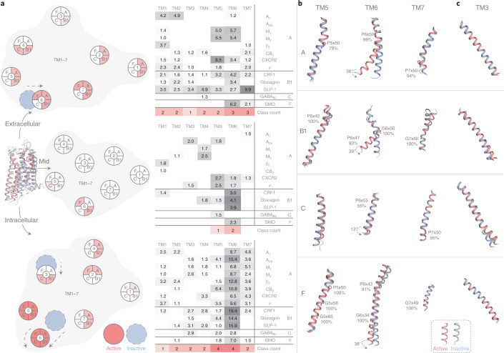
Fig. 2. Transmembrane helix movement upon activation, and universal TM3 and TM6 helix ‘macroswitches’.
a, Movements (Å) over 1.0 Å at the extracellular end, membrane mid (determined using ref. 43) and intracellular end of the transmembrane helices TM1–7 upon comparison of all available receptor inactive- and active-state structure pairs (Supplementary Table 1). Red intensity denotes the number of classes with a consensus. b, Movement and conserved hinges of TM6 and the adjacent TM5 and TM7. c, TM3 cytosolic tilt and overall rotation. b,c, GPCR class-representative inactive/active receptor structure pairs: A: β2 (refs. 44,45), B1: GLP-1 (refs. 18,22), C: GABAB246 and F: Smoothened47,48 receptors. Proline and glycine residues that increase helix plasticity are shown, along with their percentage conservation in the GPCR class.

