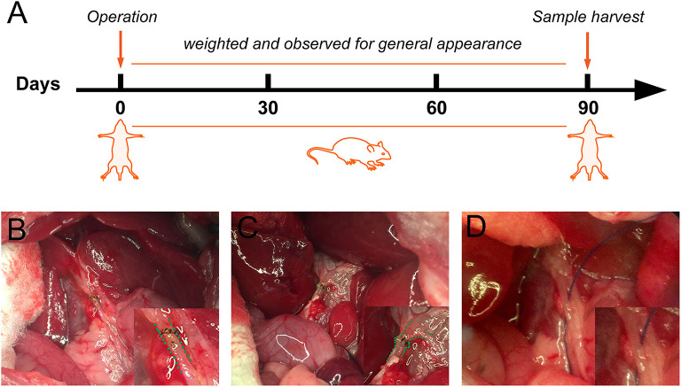
Figure 1.
Schematic representation of the experimental procedure and anastomosis methods for bile duct injured rats. (A) The flowchart of rat experiment. (B) Traditional biliary anastomosis in the control group. Common bile duct after an end-to-end suture was highlighted by a green dashed line. (C) “Fish-mouth to fish mouth” anastomosis was done in the fish mouth shaped (FMS) group. The highlighted common bile duct was reconstructed by a wider anastomosis. (D) The common bile duct was isolated in the sham group.

