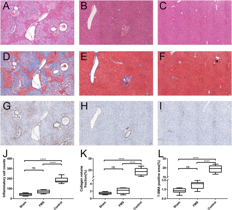
Figure 4.
The FMS reconstruction relieves the incidence of liver injury in vivo. The sections of liver tissue were stained with H&E. [(A) control group. (B) FMS group. (C) sham group], Masson [(D) control group. (E) FMS group. (F) sham group] and α-smooth muscle actin (α-SMA) antibody [(G) control group. (H) FMS group. (I) sham group]. The interstitial inflammatory cells were calculated in each group and measured (J). There were lots of inflammatory cell infiltrations in the control group, but insignificant differences between the FMS group and sham group. Collagen volume fraction (CVF) was evaluated using Image Pro-Plus 6.0, there was no statistically significant difference in the FMS group and control group (K). The α-SMA positive area in the three groups was also evaluated, similar results were seen with CVF (L). ****P < 0.0001.

