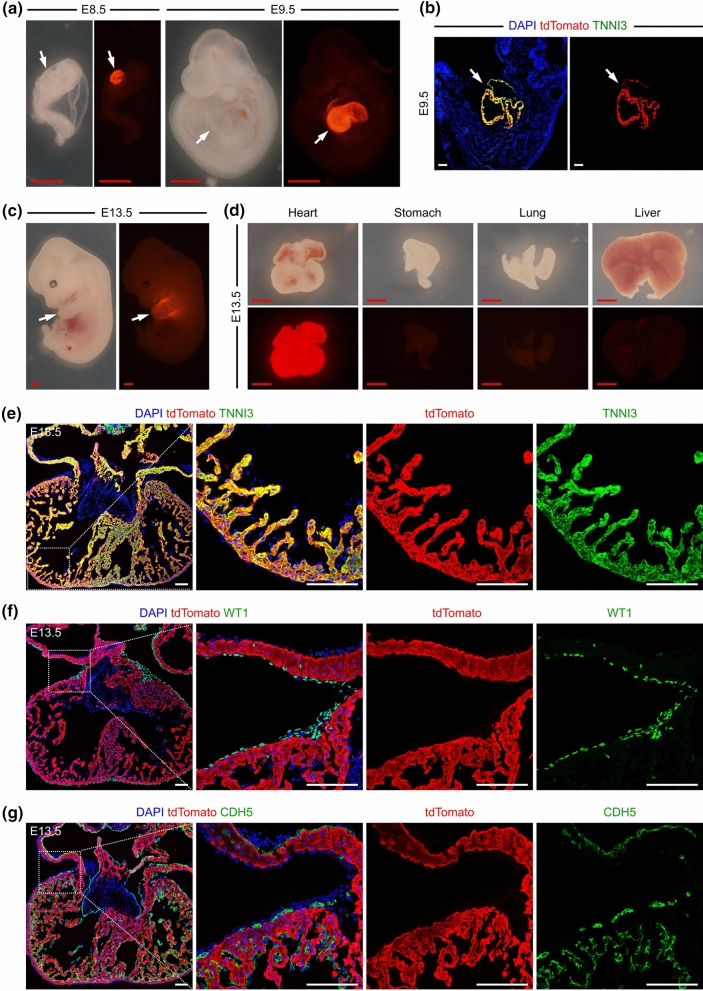
Fig. 2.
Myh6-Cre labels cardiomyocytes in embryonic hearts. (a, c) Whole-mount view of the Myh6-Cre/ + ;R26-tdTomato/ + embryos at E8.5 and E9.5. b Immunostaining for tdTomato and TNNI3 on embryonic sections at E9.5. The arrows in (a–c) indicate hearts with enriched tdTomato signals. d Whole-mount view of the organs from Myh6-Cre/ + ;R26-tdTomato/ + mice at E13.5. (e–g) Immunostaining for tdTomato and TNNI3, WT1 or CDH5 on heart sections from Myh6-Cre/ + ;R26-tdTomato/ + mice at E13.5. Boxed regions are magnified on the right. 4 embryos were examined for each embryonic stage. Red scale bars, 1 mm; White scale bars, 100 µm

