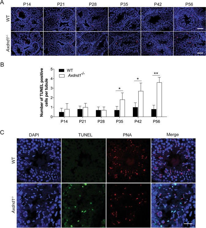
Fig. 5. Axdnd1−/− mice exhibit significantly increased apoptosis of late spermatids.
A Representative images of the apoptosis of germ cells in developmental testes were determined by TUNEL staining (red). Scale bar: 50 µm. B Quantification of the number of TUNEL positive cells in (A). Data are presented as Mean ± SEM, n = 3. *P < 0.05 by Student’s t-test. C Co-staining of TUNEL and PNA in the adult testes from WT and Axdnd1−/− mice. TUNEL (green), PNA (red), and DAPI (blue) were used to mark the apoptotic germ cells, acrosome, and nuclei, respectively. Scale bar: 20 µm.

