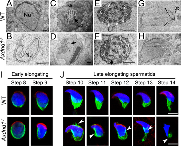
Fig. 6. Axdnd1 deletion in mice leads to abnormal spermatid development in late spermiogenesis.

A–H Transmission electronic microscopy (TEM) analysis of testicular spermatids in WT and Axdnd1−/− mice. A normal condensing sperm nucleus in WT elongating spermatids (step 8–9) is shown in (A) and an irregular-shaped sperm nucleus coupled malformed acrosome in Axdnd1−/− elongating spermatids is shown in (B). The cross-section of flagella in WT (C and E) and Axdnd1−/− (D and F) elongated spermatids are shown. Normal manchette located on the two sides of girdles of the nucleus in the WT elongating spermatid (G) and extra manchette microtubules appeared in the nucleus of Axdnd1−/− elongating spermatid. Nu, nucleus; Mt, mitochondria; Od, outer dense fiber; Bb, basal body; M, manchette. Scale bar: 1 µm in (A–B), 200 nm in (C–F), 100 nm in (G–H). I Representative co-immunofluorescent staining images of EB3 (green) with PNA (red) in early elongating spermatids (step 8–9) between WT and Axdnd1−/− are shown. J Representative co-immunofluorescent staining images of EB3 (green) with PNA (red) in late elongating spermatids (step 10–14) between WT and Axdnd1−/− are shown. Residual manchette microtubules in the acrosome, as well as rigid and disorganized manchette microtubules (labeled by white arrowheads), were observed in step 10–14 spermatids of Axdnd1−/− mice. Scale bar: 5 µm.
