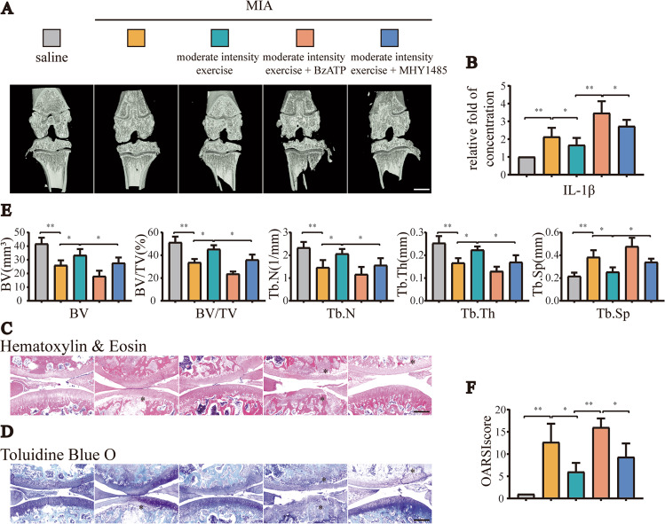
Fig. 2. Increased expression of P2X7 or inhibition of autophagy offset the anti-inflammatory effects of moderate-intensity exercise.
We set the following five groups: saline, MIA, MIA + moderate-intensity exercise, MIA + moderate-intensity exercise + BzATP, MIA + moderate-intensity exercise + MHY1485. A total of 50 rats were randomly assigned, with 10 rats in each group. A Micro-CT scanning of knee joints in each group was used to obtain imaging data for the tibial plateau and subchondral bone (scale bar: 1 mm). B ELISA was used to determine IL-1β levels in the articular cavity lavage fluid of rats in each group. C H&E staining and D TB staining were used to analyze the degree of cartilage loss and the development of inflammation. E Knee joint bone-related parameters, including BV, BV/TV, Tb.N, Tb.Th, and Tb.Sp, as evaluated by micro-CT. F OARSI scores were used to determine the development stage of OA in the knee joint tissues of rats in each group. The * in the HE and TB staining images represents the cartilage damage site. Data are presented as means ± standard deviations of at least three independent experiments. *p < 0.05, **p < 0.01.

