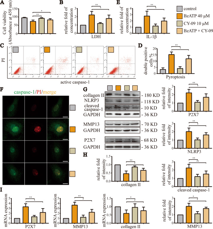
Fig. 5. Inhibition of NLRP3 alleviated OA caused by overactivation of P2X7.
A CCK-8 assays were used to detect cell viability. Absorbance was measured at a wavelength of 450 nm. B LDH release assays were used to detect the degree of cell damage. C Flow cytometry was used to detect the number and ratio of caspase-1/PI-stained cells, reflecting the severity of cell pyroptosis. D Statistical data for stained cells. E ELISA was used to detect IL-1β levels in cell culture supernatants for each group. F Cell fluorescence experiments were used to determine the fluorescence intensity and location of caspase-1/PI staining in cells, reflecting the severity of cell damage and pyroptosis (scale bar: 10 μm). G, H Western blotting and I RT-qPCR were used to detect the protein and mRNA expression levels of P2X7, NLRP3, caspase-1, MMP13, and collagen II. Data are presented as means ± standard deviations of at least three independent experiments. *p < 0.05, **p < 0.01.

