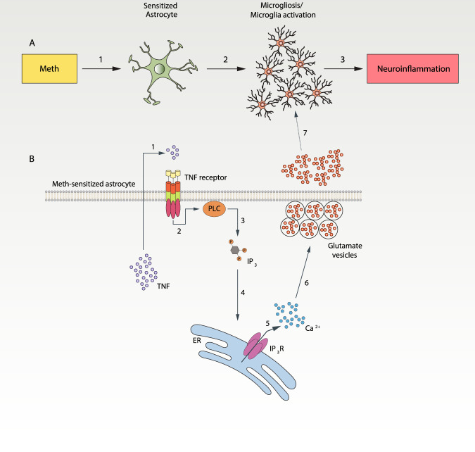
Fig. 5. Meth-induced microglia activation occurs via astrocytes.
A Exposure to Meth induces astrocytic sensitization (1). Meth-sensitized astrocytes secrete soluble factors (2) that will act on microglia causing microgliosis and microglia activation, promoting neuroinflammation (3). B In Meth-sensitized astrocytes, Meth exposure triggers the production and secretion of TNF (1). TNF acts on astrocytic TNF receptors in an autocrine manner, leading to the activation of PLC (2). TNF-induced PLC activation produces the second messenger IP3 (3) that interacts with IP3 receptors on the ER (4). Activation of IP3R2 promotes Ca2+-mobilization from the ER into the cytosol (5), consequently increasing glutamate release (6). Increased glutamate and TNF content in the extracellular milieu promotes the activation of microglia (7). Although our work focuses on the TNF-dependent effect on glutamate release, TNF released from astrocytes can interact with TNFRs present in the microglia and contribute to microglial activation observed in response to Meth. TNF tumor necrosis factor, PLC Phospholipase C, IP3 Inositol (1,3,4) phosphate, ER endoplasmic reticulum, Ca2+ calcium ions, TNFR tumor necrosis factor receptor.

