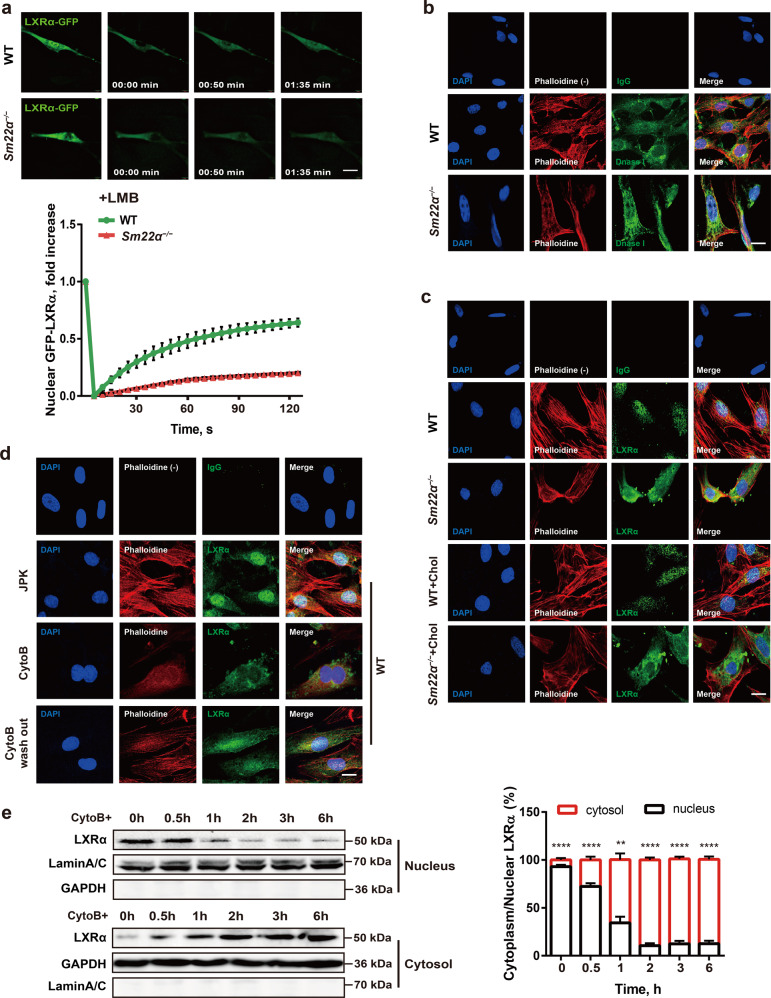
Fig. 4. Nuclear import of LXRα is regulated by actin dynamics.
a Fluorescence recovery after photobleaching (FRAP) studies with LXRα-GFP to measure nuclear import. Cells were pretreated with LMB. Decreased accumulation of nuclear fluorescence indicates a lower rate of nuclear import of LXRα-GFP in Sm22α−/− VSMCs relative to WT controls (n = 25). b Representative images of F-actin (phalloidin, red) and G-actin (DnaseI, green) in WT and Sm22α−/− VSMCs. Scale bar, 10 μm. c, d Representative images for F-actin (phalloidin, red) and LXRα (green) in WT and Sm22α−/− VSMCs with cholesterol loading or not (c) and in WT VSMCs treated with JPK, CytoB and after CytoB washout (d). Scale bars, 10 µm. e Western blot analysis of cytoplasmic and nuclear LXRα in WT VSMCs treated with CytoB at different time points (n = 6). Data and images represent at least three independent experiments. Statistical analyses, unpaired t-test and Kruskal–Wallis rank sum test. **p < 0.01; ***p < 0.001; ****p < 0.0001.

