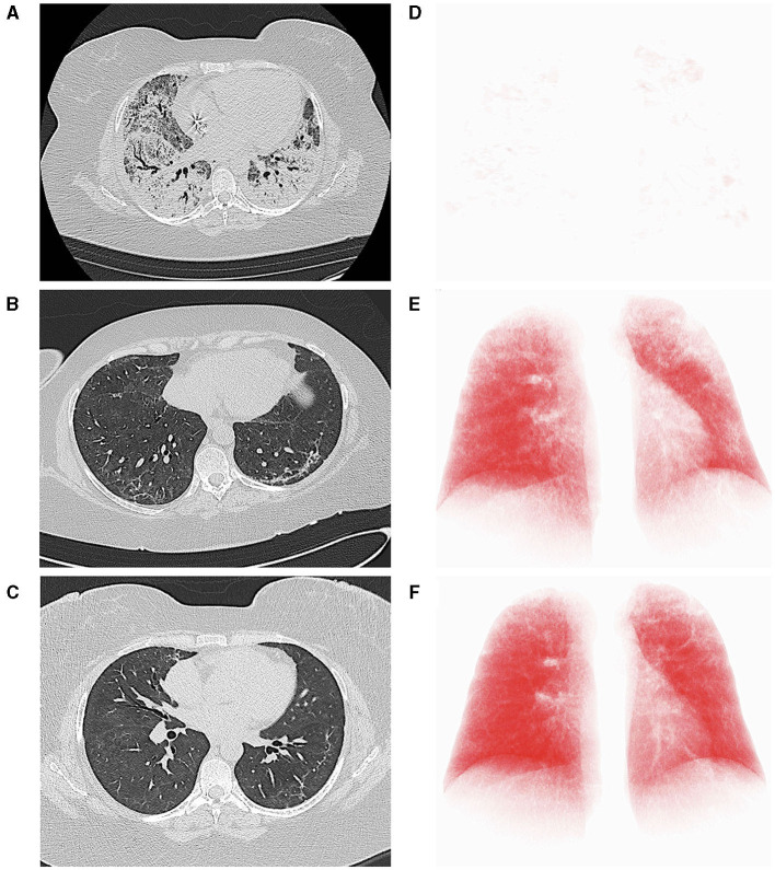
Figure 2.
Computed tomography (CT) axial scans (lung parenchyma windowing) acquired, respectively, immediately before (A), 27 (B) and 111 days (C) after the start of nintedanib, demonstrating progressive improvement of lung parenchyma and significant reduction of residual lung damage. At each time point, the axial CT image most representative of the burden of lung involvement was selected. (D–F) report the corresponding three-dimensional volume rendering images (IntelliSpace Portal v.8.0, Philips Medical Systems, Eindhoven, The Netherlands). The red volume represents the well-aerated parenchyma.

